Introduction to Tissue Types

Types of tissue coloring page – Yo, Medan peeps! Let’s dive into the amazing world of tissues – not the Kleenex kind, but the building blocks of your body! We’re talking about the different types of tissues that make you, you. It’s all about how these tissues work together to keep you functioning smoothly, from your heartbeat to your brainpower. Think of it like a super awesome team, each member with a specific role.
Our bodies are made up of four main types of animal tissues: epithelial, connective, muscle, and nervous tissue. Each type has a unique structure and function, working in harmony to keep everything running like a well-oiled machine (or maybe a really efficient warung!).
Epithelial Tissue
Epithelial tissue is like the body’s protective outer layer and lining. Think of it as the skin on your face, the lining of your stomach, or the cells that form the air sacs in your lungs. These tissues form barriers, protect underlying structures, and often have specialized functions like absorption or secretion. For example, the epithelial cells in your intestines absorb nutrients from the food you eat, while those in your salivary glands secrete saliva to help with digestion.
They’re everywhere, man! They are tightly packed together, forming sheets or layers. Some epithelial tissues are just one cell layer thick, while others are multiple layers thick, depending on their function and location.
Connective Tissue
Connective tissue is all about support and connection. It’s like the glue that holds everything together! This includes bones, cartilage, tendons, ligaments, and blood. Bones provide structural support and protection, cartilage cushions joints, tendons connect muscles to bones, ligaments connect bones to other bones, and blood transports oxygen and nutrients throughout the body. They are characterized by a matrix, which is a non-cellular substance that surrounds the cells.
The properties of the matrix vary depending on the type of connective tissue. For instance, bone matrix is hard and mineralized, whereas blood matrix is fluid.
Muscle Tissue
Muscle tissue is responsible for movement, both voluntary and involuntary. Think about flexing your biceps (voluntary) or your heart beating (involuntary). There are three types: skeletal muscle (attached to bones for movement), smooth muscle (found in internal organs like the stomach and intestines), and cardiac muscle (found only in the heart). These muscles all contain specialized proteins that allow them to contract and relax, producing movement.
Skeletal muscle is striated, meaning it has a striped appearance under a microscope. Smooth muscle is not striated and cardiac muscle has a unique striated appearance with branched cells.
Nervous Tissue
Nervous tissue is the communication network of your body. It’s like the super-fast internet connection of your system, sending and receiving signals to and from your brain and the rest of your body. This includes your brain, spinal cord, and nerves. Nervous tissue is made up of neurons (nerve cells) that transmit electrical signals and glial cells that support and protect neurons.
This rapid communication is crucial for everything from reflexes to complex thought processes.
Tissue Locations in the Human Body
Here’s a simplified view of where you can find these tissue types in your body. Remember, many organs contain multiple tissue types working together!
| Tissue Type | Location Example 1 | Location Example 2 | Location Example 3 |
|---|---|---|---|
| Epithelial | Skin | Lining of the digestive tract | Lungs |
| Connective | Bones | Cartilage | Blood |
| Muscle | Biceps | Heart | Stomach |
| Nervous | Brain | Spinal cord | Nerves |
Epithelial Tissue Coloring Page Ideas: Types Of Tissue Coloring Page
Yo, peeps! Let’s dive into some seriously fun and educational coloring page designs featuring epithelial tissues. We’re talking about those amazing cells that cover body surfaces, line body cavities, and form glands – the unsung heroes of our anatomy! Get ready to unleash your inner artist and learn something cool at the same time.Epithelial tissues come in various shapes and sizes, each with its own unique job.
These coloring pages will help visualize these differences and make learning about them a breeze. We’ll focus on three main types: squamous, cuboidal, and columnar.
Squamous Epithelial Tissue Coloring Page
Imagine a flat, tile-like surface. That’s essentially what squamous epithelium looks like. Think of it as a thin, protective layer. For our coloring page, we can represent this with tightly packed, flat cells resembling thin, slightly overlapping scales. Use a light blue for the cytoplasm and a darker blue for the nuclei, which should be small and centrally located.
Maybe add a subtle, light grey pattern to mimic the cell membrane. The overall effect should be smooth and sleek, reflecting the tissue’s function of allowing for easy passage of substances.
Cuboidal Epithelial Tissue Coloring Page
Now, picture cube-shaped cells. That’s our cuboidal epithelium! These cells are involved in secretion and absorption, and you’ll find them lining kidney tubules and glands. In our coloring page, we can depict them as neatly arranged cubes. Use a vibrant yellow for the cytoplasm and a dark orange for the round, centrally located nuclei. A light brown grid-like pattern could represent the cell boundaries, emphasizing their cubic shape and arrangement.
This design should convey a sense of structure and organization, highlighting the cells’ secretory and absorptive functions.
Columnar Epithelial Tissue Coloring Page
Lastly, let’s tackle columnar epithelium. These are tall, column-shaped cells, often with microvilli or cilia on their apical surface. They’re typically found lining the digestive tract and parts of the respiratory system. For our coloring page, we can draw these cells as elongated rectangles. Use a bright green for the cytoplasm, with darker green shading at the base of each cell to indicate depth.
The nuclei should be oval and located near the base of each cell. To show microvilli, you can add tiny, hair-like projections on the apical surface (the top) using a light purple color. This will represent the increased surface area for absorption. For cilia, use short, wavy lines of a light blue. This design should communicate the height and specialized functions of these cells, showcasing the microvilli or cilia.
Connective Tissue Coloring Page Ideas

Yo, Medan peeps! Let’s dive into the awesome world of connective tissues – the unsung heroes holding our bodies together. These coloring pages will help you visualize these crucial tissues in a fun and memorable way. We’re not just coloring; we’re building a deeper understanding of how our bodies work!Connective tissues are everywhere, from the strong bones in your frame to the flexible cartilage in your nose.
These coloring pages will focus on showcasing the unique structures of different connective tissues, highlighting both the cells and the important extracellular matrix that surrounds them. The extracellular matrix is like the scaffolding – it gives the tissue its strength, flexibility, and overall properties. Get ready to color your way to a better understanding of your body’s amazing architecture!
Bone Tissue Coloring Page
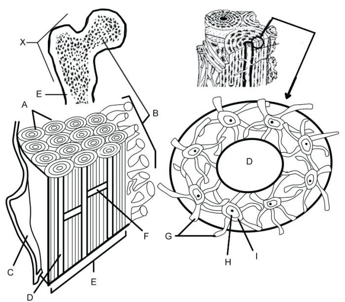
This coloring page depicts a section of compact bone tissue. Imagine a detailed view, showing the concentric rings of bone matrix (lamellae) surrounding a central canal (Haversian canal) containing blood vessels and nerves. Osteocytes, the bone cells, are nestled within small spaces called lacunae, connected to each other via tiny canals called canaliculi. The overall structure resembles a target or a tree trunk cross-section.
- Extracellular Matrix: Color the lamellae in shades of cream or beige to represent the collagen fibers and mineral deposits giving bone its strength and hardness. The Haversian canals should be a darker color, perhaps red or pink, to highlight the blood supply.
- Cell Types: Osteocytes can be colored in a light brown or tan, and they should be depicted within the lacunae. The canaliculi, connecting the lacunae, can be shown as thin, darker lines.
Cartilage Coloring Page
This coloring page will show hyaline cartilage, the most common type. Think of a smooth, glassy surface – that’s what hyaline cartilage looks like. The coloring page will display chondrocytes, the cartilage cells, residing within lacunae scattered throughout the extracellular matrix. Unlike bone, cartilage lacks blood vessels, which contributes to its slower healing rate.
- Extracellular Matrix: The extracellular matrix of hyaline cartilage is primarily composed of collagen fibers embedded in a gel-like ground substance. Color this matrix in a light blue or translucent shade to represent its smooth, flexible nature.
- Cell Types: The chondrocytes, located within the lacunae, can be colored in a light purple or blue. Show them dispersed throughout the matrix, not in organized rings like in bone.
Blood Tissue Coloring Page
This coloring page features a drop of blood, showing its cellular components. You’ll see red blood cells (erythrocytes), white blood cells (leukocytes), and platelets (thrombocytes) suspended in a fluid called plasma. The plasma is the extracellular matrix in this case. Think of it like a bustling city, with different types of cells performing their specific roles.
- Extracellular Matrix: Color the plasma in a pale yellow or straw color. It’s the liquid component carrying all the other blood cells.
- Cell Types: Red blood cells can be colored red, of course! White blood cells can be various colors (e.g., purple, green, blue) to represent the different types. Platelets can be small, dark purple dots.
Muscle Tissue Coloring Page Ideas
Yo, peeps! Let’s dive into the awesome world of muscle tissue coloring pages. We’re gonna make these pages not just fun, but also educational, showing off the cool differences between the three main muscle types. Get your crayons ready!This section details three coloring page concepts, each focusing on a different type of muscle tissue: skeletal, smooth, and cardiac.
We’ll break down the unique structures of each and how you can use color and shading to bring them to life on your coloring page. Think vibrant hues and strategic shading – it’s gonna be lit!
Skeletal Muscle Coloring Page, Types of tissue coloring page
This coloring page will showcase the long, cylindrical shape of skeletal muscle fibers, arranged in parallel bundles. The key feature to highlight is the striations – those repeating bands of light and dark. Imagine a bunch of tiny, perfectly aligned stripes running along the length of each fiber. For the coloring, use a light color for the lighter bands (perhaps a pale yellow or light blue) and a darker shade for the darker bands (maybe a deep blue or a reddish-brown).
Subtle shading, gradually darkening the color towards the center of each fiber, can give a more realistic 3D effect. You could even add a fun touch by coloring in the connective tissue that bundles the muscle fibers together, using a contrasting color.
Smooth Muscle Coloring Page
Unlike skeletal muscle, smooth muscle fibers are spindle-shaped, meaning they’re thick in the middle and taper at the ends. They lack the striations that are so prominent in skeletal muscle. Your coloring page should reflect this smooth, uniform appearance. Use a single, consistent color for the smooth muscle fibers, maybe a soft pastel shade like light green or lavender.
You can add subtle shading to give the fibers depth, but avoid any striations. To further enhance understanding, you can illustrate smooth muscle in the walls of an organ like the stomach or intestines, showing how it helps these organs move and contract.
Cardiac Muscle Coloring Page
Cardiac muscle, found only in the heart, has a unique structure. Its cells are branched and interconnected, forming a network. Like skeletal muscle, cardiac muscle exhibits striations, but these are less distinct than in skeletal muscle. The branching pattern and the intercalated discs (special junctions between cells) are important structural features to depict. Use a color palette that suggests the rhythmic pumping action of the heart—perhaps reds and oranges for the muscle fibers, with darker shading around the intercalated discs to highlight their role in coordinating contractions.
Illustrating the branching pattern with careful shading and color transitions will make this page really pop.
Nervous Tissue Coloring Page Ideas
Medan, man! Let’s get this coloring page buzzing with neurons and glial cells! We’re talking about the communication highway of your body – the nervous system. This coloring page will be a fun and educational trip into the microscopic world of nerve cells, showing how they work together to send signals all over the place. Think of it as a vibrant map of your brain’s amazing electrical network.This coloring page will depict the intricate details of neurons and their interactions, highlighting the key structural components and the process of communication between them.
It’s gonna be super cool!
Neuron Structure
The coloring page will showcase a typical neuron, clearly illustrating its three main parts: the cell body (soma), the dendrites, and the axon. The cell body will be depicted as a large, roundish structure containing the nucleus – the control center of the neuron. Branching out from the cell body will be numerous dendrites, shown as shorter, thinner extensions resembling a tree’s branches.
These will be colored differently to emphasize their role in receiving signals from other neurons. Extending from the opposite side of the cell body will be the axon, a long, slender projection that transmits signals to other neurons, muscles, or glands. We’ll make the axon a distinct color and possibly show it covered in a myelin sheath (a fatty layer that speeds up signal transmission) for extra detail.
Synaptic Communication
The coloring page will visually represent the communication between neurons at the synapse. We’ll show two neurons positioned close together, but not touching. The axon terminal of the sending neuron (presynaptic neuron) will be depicted with small vesicles (sacs containing neurotransmitters) – these will be tiny circles of a different color. The gap between the neurons, the synaptic cleft, will be clearly indicated.
The receiving neuron (postsynaptic neuron) will have its dendrites shown close to the synaptic cleft, with receptor sites that are ready to receive the neurotransmitters. The process of neurotransmitter release, diffusion across the cleft, and binding to receptors can be represented by arrows and color changes to show the signal’s journey. We can even add a little legend to explain what each color and shape represents for extra clarity.
This will bring the whole process to life, making it easily understandable.
Illustrative Examples of Tissue Types
Medan style, right? Let’s dive into some microscopic views of tissues – think of it like zooming way in on your body parts! We’ll look at examples of epithelial, connective, and muscle tissue, and how these complex structures could be simplified for a coloring page. It’s gonna be a trip!
Epithelial Tissue: Simple Squamous Epithelium
Imagine a sheet of incredibly thin, flat cells, like tiny, overlapping floor tiles. That’s simple squamous epithelium. These cells are arranged in a single layer, and their flattened shape allows for easy passage of substances. You’d see the nuclei of these cells as small, dark, slightly oval shapes, centrally located within each cell’s thin cytoplasm. The overall appearance is smooth and somewhat transparent.
For a coloring page, you could simplify this by representing each cell as a slightly elongated oval with a smaller circle inside to represent the nucleus. The coloring could be a light pastel shade to reflect the transparency.
Connective Tissue: Dense Regular Connective Tissue
Now picture a tightly packed, organized arrangement of long, parallel fibers. This is dense regular connective tissue, the kind you find in tendons and ligaments. The fibers themselves would appear as long, wavy lines running in the same direction, stained a deep pink or red. You’d see fewer cells compared to the fibers, with the cells appearing elongated and squeezed between the fibers.
These cells, fibroblasts, would be much smaller and less prominent than the fibers. To simplify this for a coloring page, focus on the parallel lines representing the collagen fibers. You could add small, simple shapes to represent the fibroblasts scattered between the lines. A simple color scheme of brown or red for the fibers and a lighter color for the cells would work well.
Muscle Tissue: Skeletal Muscle Tissue
Think of long, cylindrical fibers, arranged in parallel bundles. This is skeletal muscle tissue. Each fiber is multi-nucleated, meaning you’d see many small, oval nuclei at the periphery (edges) of each fiber. The fibers themselves show a distinct striated (striped) appearance due to the arrangement of contractile proteins – these stripes would appear as alternating light and dark bands running across the fibers.
For a coloring page, represent each muscle fiber as a long, slightly irregular rectangle, with multiple small circles at the edges representing the nuclei. The stripes could be simplified to alternating thick and thin lines across each fiber. Using a combination of red and pink for the muscle fibers would create a realistic effect.
FAQ Compilation
What age groups are these coloring pages suitable for?
These coloring pages can be adapted for various age groups. Simpler designs with fewer details work well for younger children (elementary school), while older children (middle school) can handle more intricate designs and labels.
Can I use these coloring pages for educational purposes in a classroom?
Absolutely! These coloring pages are a fantastic tool for making biology lessons more engaging and memorable. They’re a great way to reinforce learning about different tissue types in a fun and creative way.
Where can I find printable versions of these coloring pages?
The provided Artikel details the
-design* of the coloring pages. You would need to create the actual printable versions using graphic design software or by hand.
Are there any specific color recommendations for each tissue type?
The Artikel suggests using color palettes that are both visually appealing and informative. For example, you might use shades of pink and red for muscle tissue to represent blood flow, or different shades of blue for connective tissue to reflect the different types.
